



Description
Product Overview
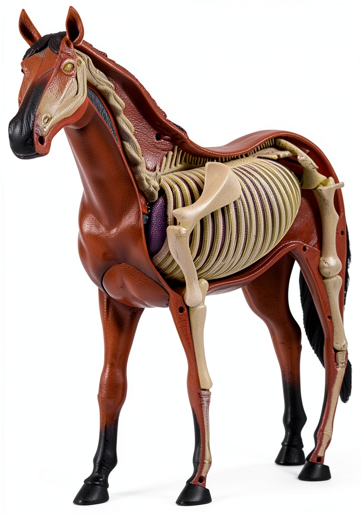
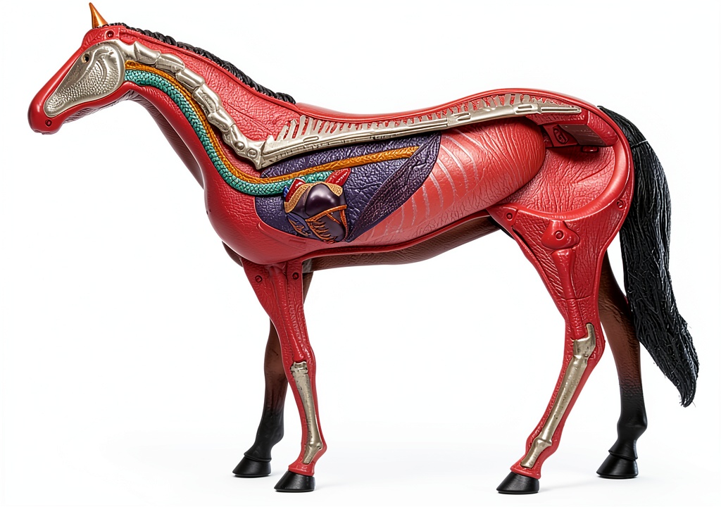
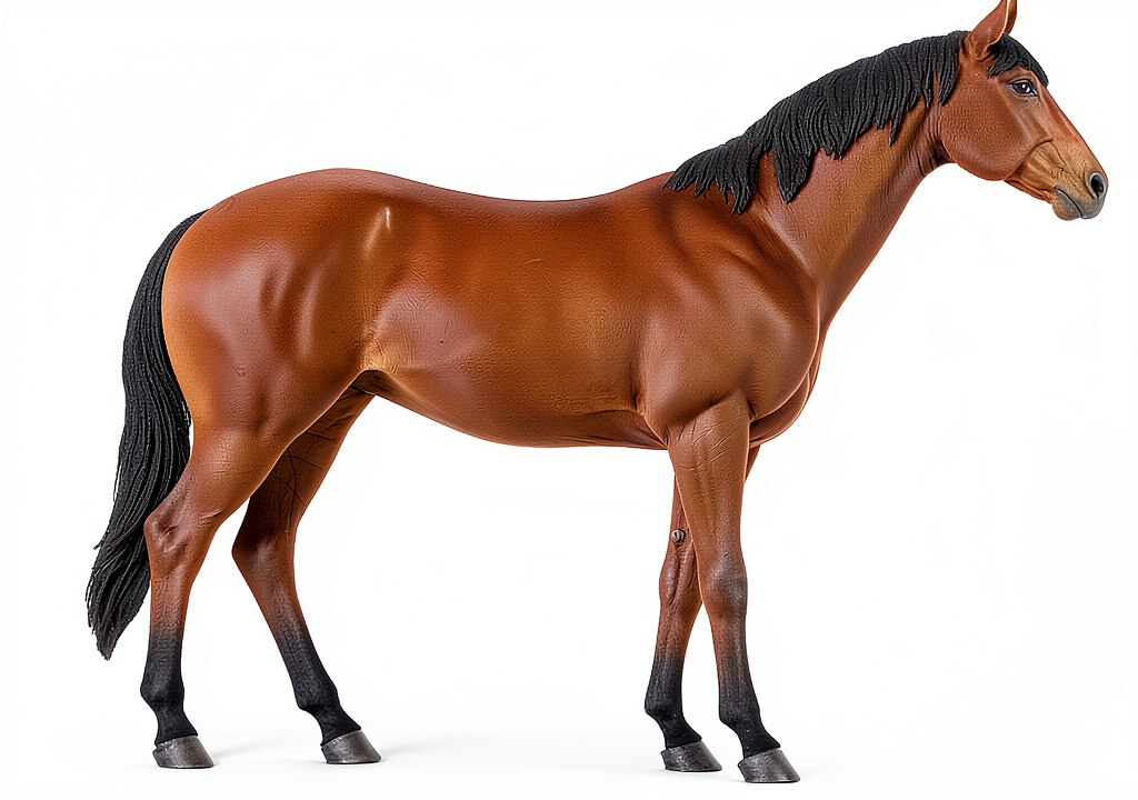
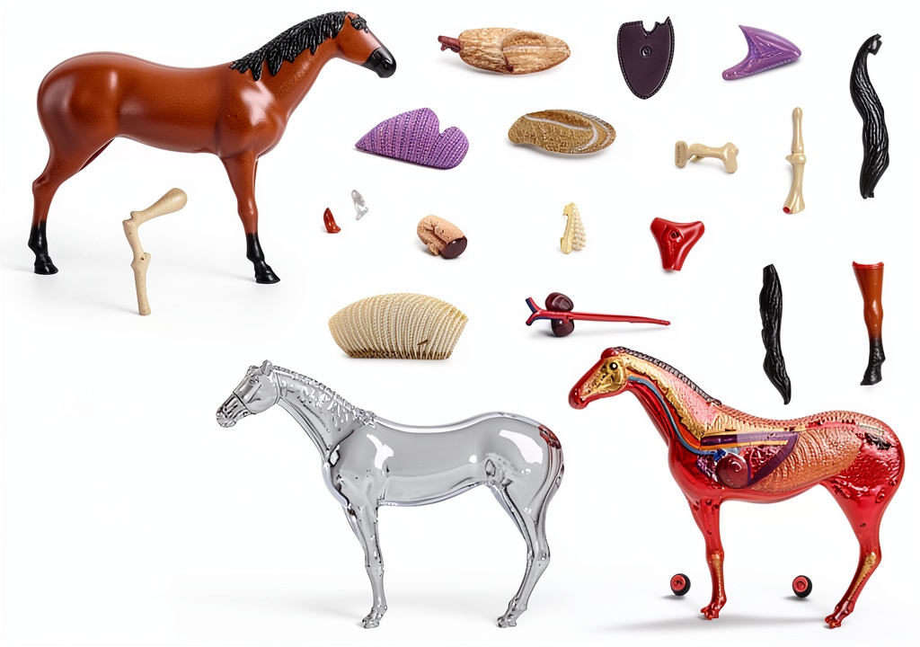
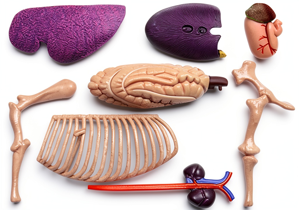
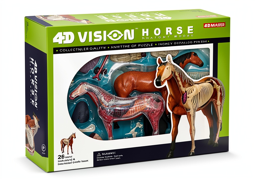
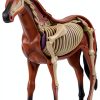
The 4D Horse Anatomy Model from SORECI is a meticulously engineered educational tool that replicates the complete equine skeletal system in twenty‑six detachable parts. Crafted from high‑impact, non‑toxic polymer, each bone is sculpted to exact anatomical proportions, providing a realistic tactile experience for students, veterinarians, and animal enthusiasts alike. Every component is clearly labeled with durable lettering, allowing users to quickly identify the scapula, femur, vertebrae and other critical structures without the need for additional reference materials. The modular design enables effortless assembly and disassembly, making it ideal for classroom demonstrations, laboratory labs, and hands‑on training sessions where repeated handling is required. With its sturdy construction and precise detailing, the model serves as a long‑lasting visual aid that supports a wide range of curricula, from basic anatomy introductions to advanced veterinary pathology studies.
The model’s 4‑dimensional representation emphasizes spatial relationships between bones, helping learners visualize how the forelimb and hindlimb connect to the axial skeleton. By physically manipulating each segment, users gain insight into joint articulation, load‑bearing mechanics, and common injury sites that are essential knowledge for equine health professionals. The inclusion of a comprehensive parts list and an instructional guide further streamlines the learning process, ensuring that educators can integrate the model into lesson plans with minimal preparation time. Whether displayed on a laboratory bench or incorporated into interactive workshops, the 4D Horse Anatomy Model enhances comprehension, retention, and confidence in anatomical identification.
The model’s dimensions are carefully calibrated to reflect the true scale of an adult horse, with the full assembled length measuring approximately 150 centimeters, the height at the withers around 120 centimeters, and the overall weight of the set near 8 kilograms. This realistic sizing allows learners to appreciate the proportional relationships between the skull, rib cage, and limb bones, facilitating a deeper understanding of biomechanics and locomotion. The color‑coded labeling system further enhances clarity, assigning distinct hues to major bone groups such as the axial, appendicular, and cranial sections, thereby simplifying the learning curve for beginners.
In addition to its primary educational function, the 4D Horse Anatomy Model serves as an effective visual aid for presentations, research posters, and veterinary conferences. Its sturdy base and optional mounting brackets enable secure display on podiums or exhibition tables, while the modular components can be arranged to illustrate specific anatomical concepts or comparative anatomy with other species. The model’s versatility makes it a valuable addition to any animal science curriculum, supporting interdisciplinary teaching that bridges anatomy, physiology, and clinical practice.
Research indicates that tactile learning tools like the 4D Horse Anatomy Model significantly improve retention rates compared to purely visual or textual instruction. By engaging multiple senses, students develop stronger neural connections related to anatomical knowledge, which translates into higher performance in practical examinations and clinical assessments. Institutions that have incorporated the model into their curricula report measurable gains in student confidence and competency, underscoring the model’s role as a catalyst for academic excellence.
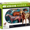
![[Product front view showing all components]](https://www.newlookshop.store/wp-content/uploads/2026/07/6b614feb45b44fcd899ab23d4a7e28ad.jpg)
Usage
In veterinary training programs, the model is employed to demonstrate fracture patterns, surgical approaches, and prosthetic fitting techniques. Instructors can simulate real‑world scenarios by removing specific bones, highlighting pathological changes, and discussing treatment options in a controlled environment. The detachable nature of the specimen also allows students to practice assembly skills, reinforcing their understanding of anatomical orientation and spatial reasoning.
Beyond professional education, the model is a valuable resource for pet clinics that wish to educate owners about equine musculoskeletal health. By showing the exact location of a sore joint or a broken bone, veterinarians can communicate treatment plans more effectively, fostering trust and compliance. Hobbyists and horse owners who are passionate about anatomy can also benefit from the hands‑on experience, turning complex concepts into accessible, visual learning.
During hands‑on workshops, instructors can challenge students to assemble the skeleton within a timed exercise, promoting teamwork, problem‑solving, and retention of anatomical terminology. The model also integrates seamlessly with digital learning platforms; educators can pair the physical specimen with augmented reality (AR) applications that overlay additional information, such as muscle attachments or vascular pathways, creating a hybrid learning environment that caters to diverse learning styles.
Equine rehabilitation centers often use the model to illustrate therapeutic protocols to owners, demonstrating how splints, casts, and supportive bandages interact with the underlying bone structure. By visualizing the exact location of a fracture or joint disease, clients gain confidence in the recommended treatment plan, leading to improved compliance and better outcomes for the animal.
Why Choose Us
SORECI’s commitment to quality is reflected in the rigorous manufacturing standards applied to every component of the horse anatomy model. The polymer material is UV‑stable, impact‑resistant, and free from hazardous chemicals, guaranteeing safety for repeated handling in busy classroom settings. Each piece undergoes precise CNC machining and hand‑finishing to achieve smooth edges and accurate anatomical contours, reducing the risk of injury and ensuring a professional appearance.
Customer support is a cornerstone of the SORECI experience. Buyers receive a detailed assembly manual, a digital reference guide, and access to a dedicated support team ready to answer technical questions or provide replacement parts if needed. This after‑sales service ensures that the educational investment remains functional and relevant throughout years of use, delivering lasting value to institutions and individuals alike.
SORECI’s design process incorporates feedback from veterinary educators, clinicians, and students to continuously refine the model’s accuracy and usability. The company conducts rigorous testing cycles, including repeated assembly‑disassembly trials, to verify that each joint maintains its integrity over thousands of cycles. This user‑centered approach ensures that the final product meets the practical demands of busy educational environments.
The environmental impact of the model is minimized through the use of recyclable polymer blends and packaging made from post‑consumer recycled paper. SORECI adheres to ISO 14001 environmental management standards, demonstrating a commitment to sustainability that resonates with institutions seeking eco‑friendly teaching resources. By choosing this model, educators not only enhance learning outcomes but also support responsible manufacturing practices.
Key Features
- Twenty‑six precisely molded bone pieces replicate the full equine skeleton, offering a complete visual and tactile reference for anatomy studies.
- Detachable design with secure interlocking joints allows easy assembly, disassembly, and transport, making the model suitable for classroom demonstrations and field workshops.
- Each component is clearly labeled with durable, fade‑resistant lettering, enabling quick identification of anatomical structures without additional reference materials.
- Constructed from high‑impact, non‑toxic polymer that resists cracking and UV degradation, ensuring long‑term durability even with frequent handling.
- Comes with a comprehensive instruction booklet and dedicated customer support, providing guidance for setup and answering any post‑purchase inquiries.
FAQ
What age or education level is the model appropriate for?
The 4D Horse Anatomy Model is designed for a wide range of learners, from high‑school biology students to graduate‑level veterinary trainees. Its clear labeling and modular construction make it accessible to beginners, while the detailed anatomical accuracy satisfies the needs of advanced professionals seeking a hands‑on reference for complex procedures.
Can the model be used to demonstrate specific injuries or conditions?
Yes, the detachable nature of the model allows educators to remove or reposition individual bones to illustrate fractures, dislocations, and pathological deformities. Instructors can add supplemental accessories such as mock casts or splints to show treatment techniques, providing a realistic simulation of clinical scenarios.
How does the model compare to other animal skeleton kits on the market?
SORECI’s horse anatomy model stands out due to its 26‑part construction, high‑impact polymer material, and color‑coded labeling system. While many kits offer basic bone replicas, this model delivers superior durability, precise anatomical detail, and comprehensive support resources, making it a more effective educational investment.
What warranty or after‑sales service does SORECI provide?
SORECI offers a one‑year limited warranty covering manufacturing defects and includes a responsive customer service team that assists with assembly questions, part replacements, and technical support. Users can contact the support portal for prompt assistance, ensuring the model remains a reliable teaching tool throughout its lifespan.







































Reviews
There are no reviews yet.